
Es befinden sich keine Produkte im Warenkorb.
24+ Months, Apicoectomy, Augma Bone Cement Academy, Bone Cement Expert, Clinical Cases, Clinical Indication, Clinician, Cyst Enucleation, Dental Notation, Images, Lower Right Molar, Media, Post-Op Period
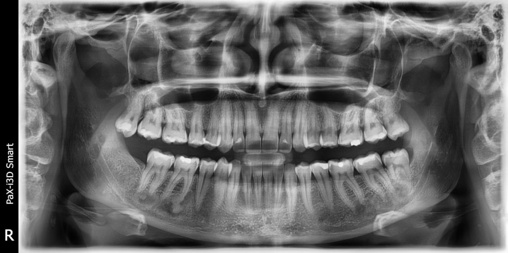
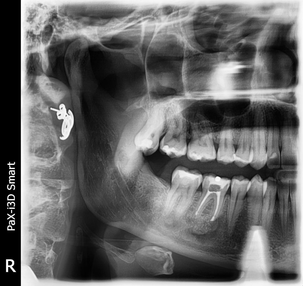
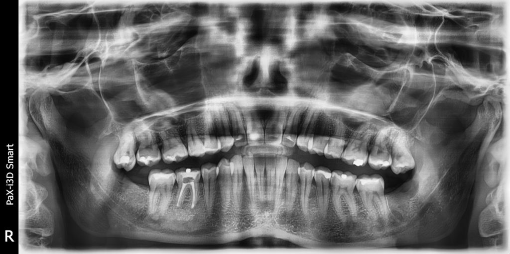
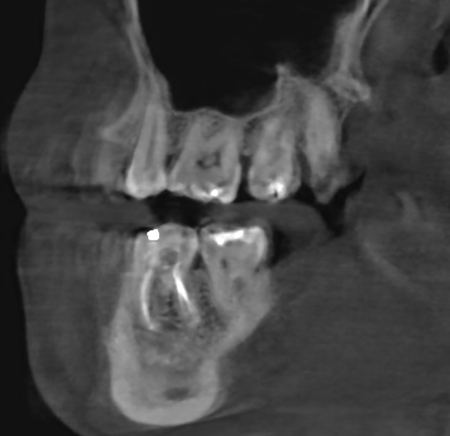
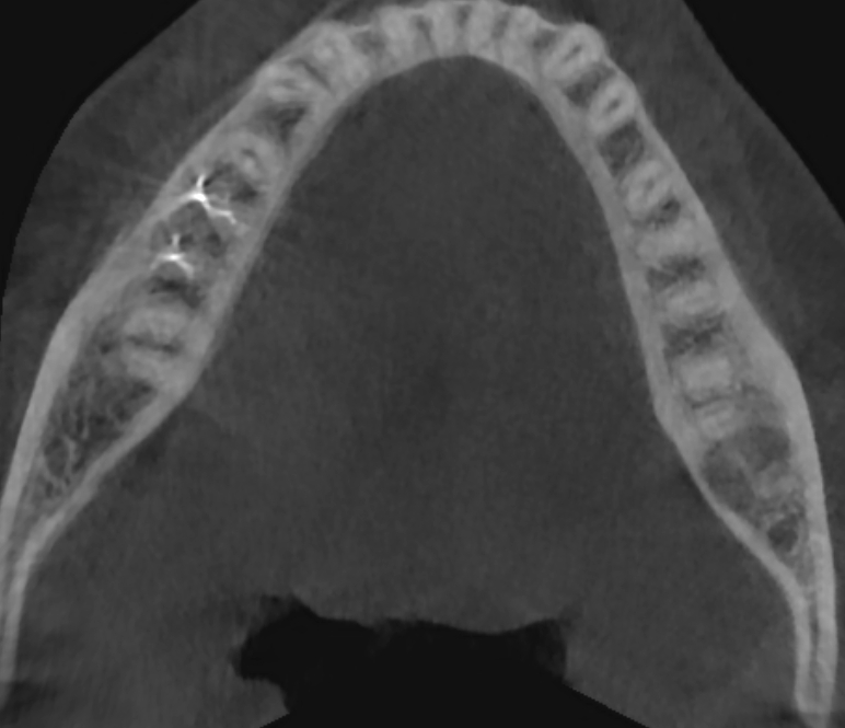
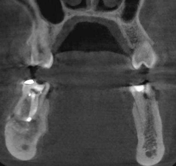
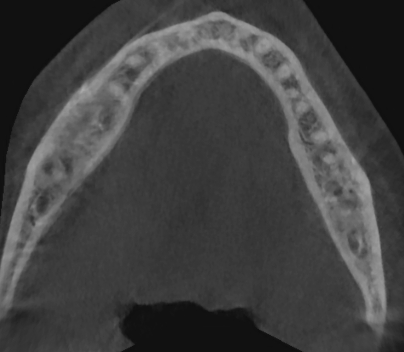
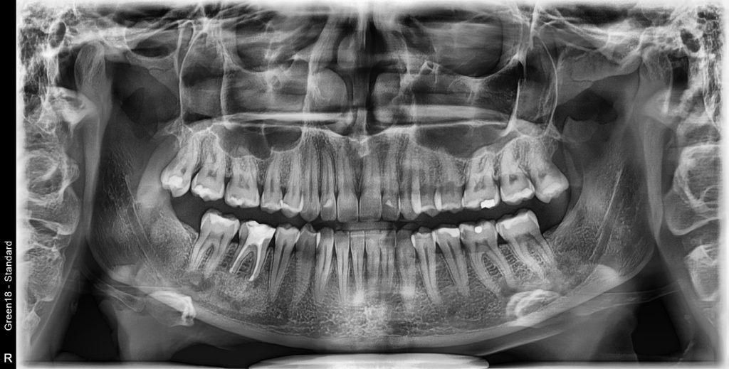
Central Odontogenic Fibroma & Central Osteoma of The Mandible
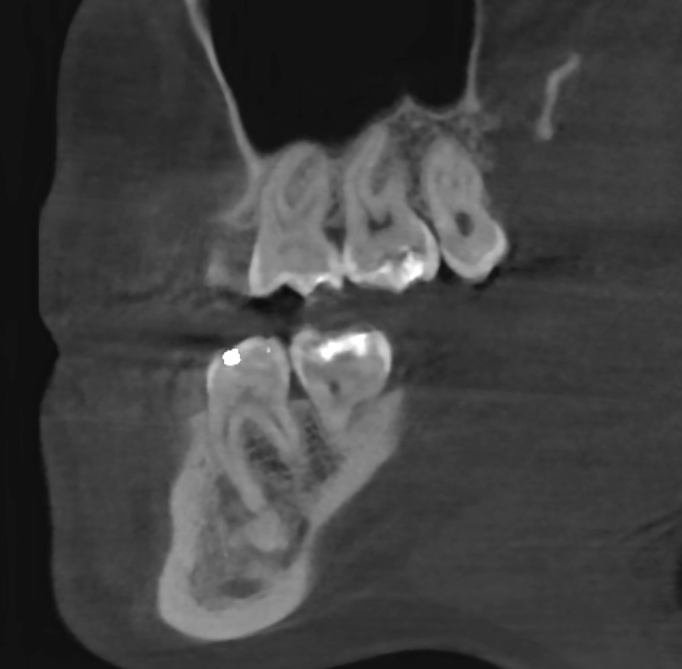
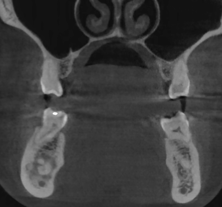
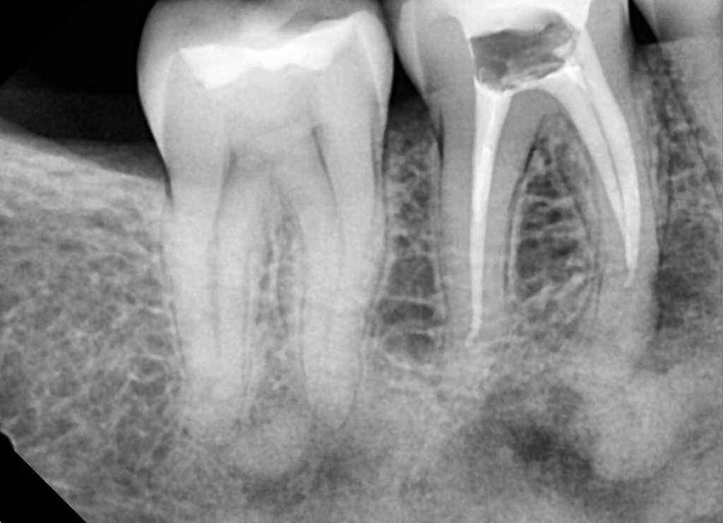
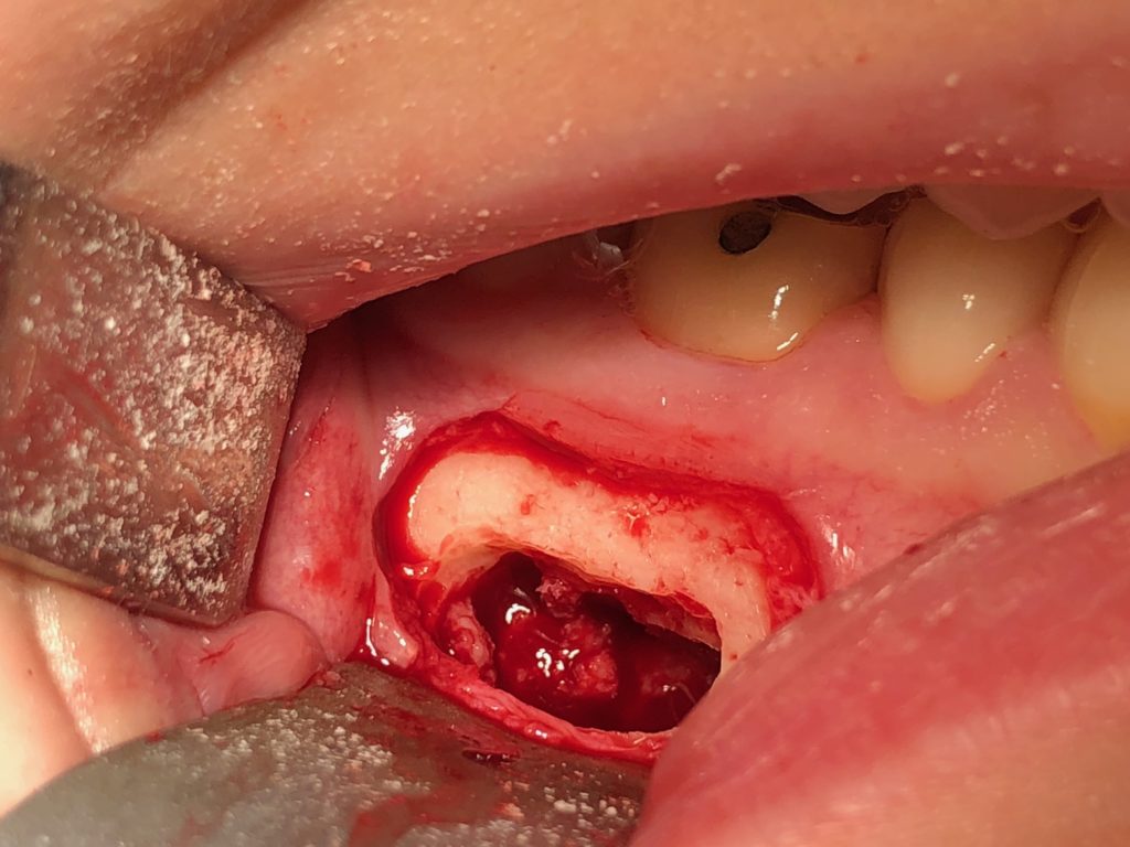
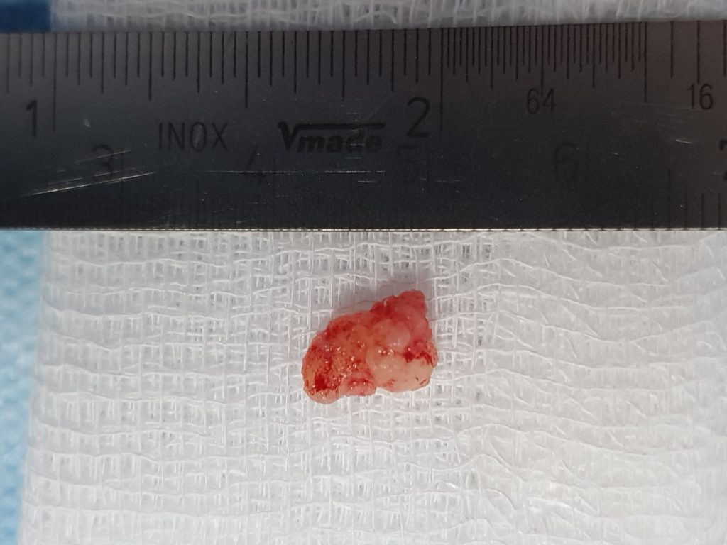
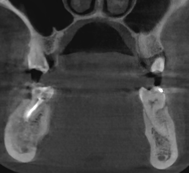
Healthy Female, age 29 years. The tumor was developed without any clinical symptoms for about 2 years. Maybe changes at the cellular level led to pulpitis and second to pulp necrosis #46 (30). The endodontic treatment was not carried out correctly, due to significant obliteration of the root canals #46 (30). A revision of the area #46 (30) was made, the tumor was removed (highly mineralized part similar to osteoma and granulation tissue from area around). We made a root resections of tooth #46 (30) with retrograde filling of the root canals using MTA. We filled the bone defect with Bond Apatite (1 cc).
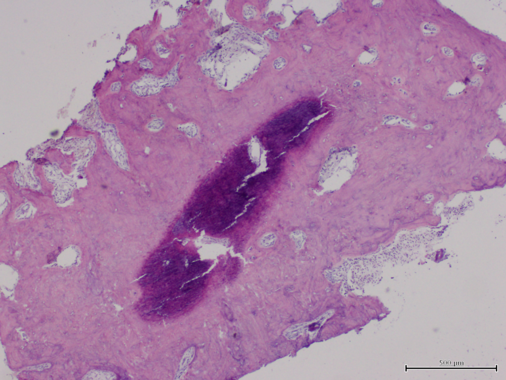
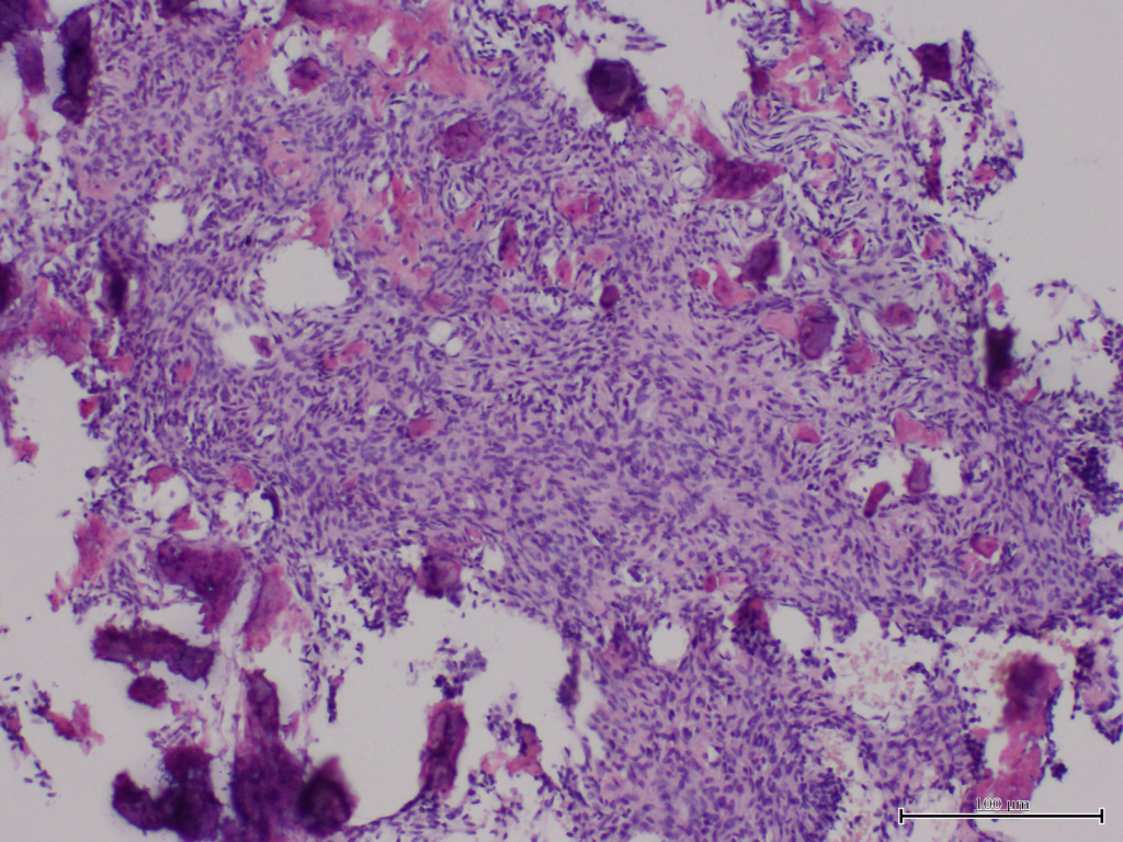
Histopathological study showed the presence of a odontogenic fibroma structure in granulation tissue and osteoma in mineralized part. We observed a good healing of bone and soft tissue in 2 years months follow-up.